Recommended Ultrasound Terminology
M
macrocirculation (tumor)
Major intranodular vessels seen as discrete hyperechoic (hyperenhanced) vessels on contrast-enhanced scans.
magnetic resonance-guided focused ultrasound
A technology that combines magnetic resonance imaging to guide focused ultrasound energy for the treatment of deep tissue.
main beam
Region of strong acoustic pressure or response centered on the beam axis (see Figure 10).
_page_11.png?sfvrsn=6387ddb_4)
Maltese cross artifact (elastography)
A fanning crosslike pattern of strain concentrations and dilutions that surrounds a circular or spherical inclusion in a medium.
manual scanning
See freehand scanning.
marginal enhancement
Similar to rim enhancement but with a rim particularly irregular and thick. A feature of hypovascular metastasis.
mass density
Amount of mass of a material per unit of volume. The unit is kilogram per cubic meter (kg/m3).
matched filtering
In signal processing, the practice of correlating a signal with a known template to detect the presence of the template in the signal. Matched filters are commonly used after coded excitation or chirp transmit pulses to produce short-duration high-amplitude echo signals with a high signal-to-noise ratio from these pulses. The filter template is derived from the code or chirp pulse.
matching layer
One or more layers of material applied to the radiating surface of a transducer with the intent of optimizing the acoustic impedance match between the transducer material and the subject. Energy transmission is optimized by choosing the impedance of the matching layer(s) to be the geometric mean(s) of the impedances of the adjoining media and the thickness of the matching layer(s) to be one-quarter wavelength in each layer.
matrix
A rectangular arrangement of elements or numbers in columns and rows.
matrix array
Transducer containing a 2-dimensional array. In general, these arrays can be other shapes than rectangular and not completely filled in as in a sparse array.
matrix element
Array element in a matrix array.
Maxwell model
A viscoelastic model of a material that can be represented by a dashpot η and a purely elastic spring E connected in series, as shown in the diagram below.

Related terms: Burgers model, dashpot, elastic spring, Kelvin model, Kelvin-Voigt model, standard linear viscoelastic model, Voigt model
mean peak cycle acoustic pressure
The arithmetic mean of the maximum positive and modulus of the maximum negative instantaneous acoustic pressure during a pulse peak cycle. The unit is pascal (Pa).
mechanical coupler
Device of predetermined mechanical impedance used for the calibration of a bone conduction vibrator or bone vibrator in conjunction with a calibrated microphone adapted to measure the pressure developed within the cavity.
mechanical impedance
Quotient of a force applied to a linear mechanical system, by the resulting velocity in the direction of the force, at its point of application. Note: In the case of torsional mechanical impedance, the words “force” and “velocity” are replaced by “torque” and “angular velocity.”
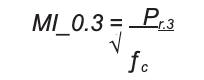
mechanical index (MI)
A dimensionless number used to represent the likelihood of an ultrasound-induced bioeffect caused by a mechanical process such as cavitation. It is equal to the ratio of the maximum negative derated negative pressure maximum amplitude, Pr.3 to the square root of the frequency in megahertz, ƒc.
mechanical reactance
Imaginary part of mechanical impedance.
mechanical resistance
Real part of mechanical impedance.
mechanical sector scanner
Scanning device in which the transducer is mechanically rotated, or pivoted, producing a fan-shaped movement of the ultrasound beam.
mechanically swept scanning (3-dimensional)
Acquiring a volume of echo information through motorized movement of the transducer to control the positions of the acquisition planes.
Related term: Contrast with electronically swept scanning
medical diagnostic ultrasonic system
The combination of the ultrasound instrument console and associated transducer assembly(ies).
medium
Any material through which a sound wave travels.
microbeamformer
A beamforming method by which fine delays are applied to subgroups of (2D) array elements and summed prior to the application of coarser delays to each subgroup.
microbubble
A small gas bubble, usually with a diameter in the 1- to 20-micrometer (µm) range. May be used as an ultrasound contrast agent.
microbubble cavitation imaging
An ultrasound imaging technique used to visualize cavitation (inertial or noninertial).
microcirculation
The system of tiny (<100 micrometer) blood vessels, including capillaries, venules, and arterioles, that perfuse the body's tissues.
microcirculation (tumor)
The network of tiny blood vessels (capillaries, arterioles, and venules) that supply blood to a tumor, developing as a consequence of neoangiogenesis. Unlike normal tissue vasculature, tumor microcirculation is often abnormal in structure and function. These vessels have a diameter of 2 to 5 µm, which is below the resolution threshold of all in vivo imaging modalities, contrast-enhanced sonography included. Contrast-enhanced grayscale sonography can detect a signal from small vessels below the resolution of unenhanced sonography and Doppler techniques. Contrast-enhanced grayscale sonography allows demonstration of tumor enhancement, but it should be always remembered that this only indirectly reflects the tumor microcirculation.
micrometer
A unit of length equal to 1 millionth of a meter, formerly called a micron. The abbreviation is µm. Also, a device for the precise measurement of small distances, typically within ±1 µm.
microparticle
A solid or liquid particle with a diameter in the micrometer range, typically 1 to 10 µm. May be used as an ultrasound contrast agent or for drug delivery.
microphone
Electroacoustical transducer by which electrical signals are obtained from acoustical oscillations.
microspheres
Small spherical bodies comprising solid, liquid, or gas, or some combination of these, usually with diameters measuring several micrometers. Microspheres may be used to enhance diagnostic imaging, to promote cavitation for therapeutic purposes, to deliver drugs or genetic material to specific locations, or for other medical reasons.
microstreaming
Small-scale eddy currents or flow in the neighborhood of ultrasonically driven oscillating bubbles.
microvascular imaging
A technique for improving depiction of small capillary vessels with a low concentration of a contrast medium.
mirror image artifact (Doppler)
The appearance of Doppler spectral components on the “wrong” side of the zero-flow baseline, so that flow spectra in both directions are mirror images of each other. Caused by a Doppler angle of 90º, by technical deficiencies in the Doppler demodulator, or by overload of the equipment.
mirror image artifact (imaging)
A multiple-path reflection artifact in which the sonographic image of a structure is duplicated in a different location and appears as a mirror image of the original (see Figure 21).
_page_22.png?sfvrsn=2c43e0f5_4)
mirroring
See mirror image artifact (Doppler).
mixer
A device that combines two or more signals. In ultrasound instrumentation, mixers multiply two signals to obtain one signal, which contains both the sum and the difference of the two input frequencies.
M-mode (motion mode)
A method of display in which the tissue interface position is displayed along one axis and slow time (comparable to the pace of the pulse repetition period) is displayed along the second axis. The M-mode is used frequently to display echocardiographic data in which heart wall motion and valve motion are displayed as functions of time.
mode conversion
A change of one type of acoustic wave to another. For example, the change from a longitudinal wave to a transverse wave (or a shear wave), and vice versa, when an acoustic wave traverses a boundary between two objects with different material properties.
mode of oscillation
See mode of vibration.
mode of vibration
A state of motion of a freely vibrating system (not driven) in which all moving parts of the system are oscillating in phase with the same frequency. A mechanical system usually can exhibit many such states of motion, each with its own frequency. The fundamental and harmonic frequencies of a violin string correspond to such a mode of vibration.
modulus
Magnitude of a quantity, the absolute value. An additional meaning is the ratio of stress to strain in an elastic or a viscoelastic medium, as in Young’s modulus.
modulus of rigidity
See shear modulus.
molecular imaging
A class of noninvasive imaging methods to visualize and characterize both normal and pathologic processes with a living organism at the cellular and subcellular levels.
molecular ultrasound imaging
An ultrasound-based imaging method that uses a target-specific microbubble to noninvasively locate and characterize both normal and pathologic processes within a living organism at the cellular and subcellular levels.
See molecular imaging.
monochromatic
Having a single frequency (from analogy with light).
monopole
See point source of sound.
monostable multivibrator
An electrical device or circuit that produces a second pulse at a known delay following the application of a triggering pulse.
mosaiclike enhancement
Term derived from computed tomographic terminology to indicate patchy enhancement. A type of inhomogeneous enhancement.
ms–1
Abbreviation for meters per second.
multi-D array
A transducer array in which transducer elements are arranged in multiple rows.
multielement transducer
A configuration of two or more transducer elements within dependent electrical addressability (see Figures 19 and 27).
_page_20.png?sfvrsn=69539fe1_6)
_page_28.png?sfvrsn=b40dff6d_3)
multifocusing
Ability of scanners employing array transducers to position the transmission or reception focus by software control or to use more than one transmitted focus at a reduced frame rate.
multigate pulsed Doppler system
Pulsed Doppler system enabling velocity measurement at several depths simultaneously.
multiplanar
Refers to or consists of several orientations. In 3-dimensional imaging, it refers to the simultaneous display of two or more orthogonal planes.
multipoint spectral Doppler
See ultrafast Doppler, ultrafast ultrasound techniques