1960s
Under the presidency of Carrie Chapman, MD (1963-1965), the first and, to date, only female AIUM president, the AIUM's official constitution and bylaws were written.
In 1964, an important turning point for the AIUM occurred. The AIUM broadened its interests, which had been focused primarily on the application of ultrasound in physical medicine, to include the use of ultrasound in all aspects of medicine - both diagnostic and therapeutic - which would eliminate the need to form 2 separate organizations. This marked the point at which AIUM membership began to expand rapidly.

Carlos Bustamente Ruiz, MD, was the AIUM's fifth president from 1965 to 1966. It was Dr Ruiz who, along with Drs Aldes and Chapman, initiated the first AIUM Pan-American meeting in Lima, Peru, in 1965.
During the AIUM's early years, Virginia Krause served as executive secretary and later was appointed to serve as administrative assistant to the Executive Board. Through her enthusiasm and devotion, Ms Krause was able to increase membership greatly and secure the first paying commercial exhibitors for the AIUM's 1964 meeting in Boston, Massachusetts.
We all know that ultrasonic therapy, combined with special fields of research, holds extraordinary and optimistic implications for even greater progress of ultrasound as a therapeutic, surgical, and diagnostic tool.
-Carrie Chapman, MD
 William J. Fry
In 1968, the AIUM became known as the American Institute of Ultrasound in Medicine rather than the American Institute of Ultrasonics in Medicine.
- Offering educational activities to acquaint those just entering the field of ultrasonics with the fundamentals of physical principles and the rationale for clinical or research use;
- Bringing together the users and developers of ultrasonics equipment to facilitate conceptual breakthroughs into working prototypes and eventually into useful clinical tools;
- Sponsoring the development of uniformly understood and accepted terminology to promote meaningful dialogue among various disciplines; and
- Initiating an equipment standardization program to permit precise determination of the acoustic parameters used to obtain sonograms and the dosage levels used in therapeutic applications.
If ultrasound is going to continue at the current rate of expansion, we need to set up training programs on a solid academic base of excellence.
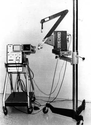
The first commercial model of a portable compound contact scanner was marketed by Physionic Engineering, Inc, as the Porta-arm Scanner
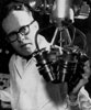
J. Stauffer Lehman, MD, scans a patient at his laboratory at Hahnemann Hospital in Philadelphia, Pennsylvania, in the early 1960s. Dr Lehman's laboratory was an important center for work in the clinical application of ultrasound in the diagnosis of abdominal and pelvic diseases in the United States.
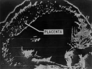
Bistable 2D Imaging
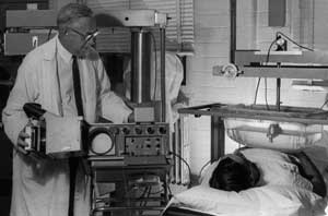
Bistable means having 2 possible states: "white or black." The image shown above was produced in 1963 using the Physionic Engineering, Inc, Porta-arm Scanner by Horace Thompson, MD, and Kenneth Gottesfeld, MD. The obstetricians were associated with the clinical ultrasound program under Joseph Holmes, MD, at the University of Colorado Medical Center. The image shows an anteriorly placed placenta.
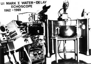
Dr George Kossoff and his coworkers at the Australian Department of Health designed ultrasound machines like the "UI Mark I" that were used clinically in the 1960s.
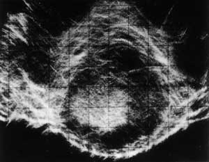
Gray Scale 2D Imaging
Gray scale is a display technique in which echo amplitude or intensity information is recorded as variations in brightness (shades of gray). True gray scaling evolved from the work of the Kossoff group. The image shown above is an early gray scale image of a placenta produced by George Kossoff, DscEng, and William Garrett, MD.