1950s
In the summer of 1951, 24 physicians attending the American Congress of Physical Medicine and Rehabilitation in Denver, Colorado, found a common interest in the validity of ultrasonic energy as a medical tool.
 Cecil J. Birtcher
Mr Cecil Birtcher, president of the Birtcher Corporation and one of the AIUM's founders, encouraged this group of ultrasound pioneers and provided them with ultrasonic instruments with which to conduct research and carry out experiments. The Birtcher Corporation assumed most of the financial obligations of the AIUM during the AIUM's early years.

 Disraeli Kobak, MD and John Aldes, MD
Disraeli Kobak, MD, the AIUM's first president, along with Mr Birtcher, organized the first annual meeting of this ultrasound group, held in New York in September 1952. These enthusiasts continued to meet and experiment with ultrasonics each year. This group evolved into the American Institute of Ultrasonics in Medicine, which later became the American Institute of Ultrasound in Medicine. John Aldes, MD, an instrumental figure in the AIUM's early days, served as the AIUM's first secretary and then as the first executive director.
In 1951, there was a growing interest in ultrasound, although how it worked was pretty much a mystery.
- Jerome Gersten, MD
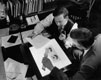
John Julian Wild, MD, PhD (left), and John Reid, PhD, compare tumor and normal echographic traces.
At the AIUM's 1955 meeting in Detroit, Michigan, John Julian Wild, MD, PhD, and John Reid, PhD, presented the first paper on ultrasound used for medical diagnostic purposes, entitled "Echographic Tissue Diagnosis."
The AIUM's focus was primarily on the use of ultrasound for therapeutic purposes, although more research on its diagnostic uses was being introduced.
As we review the past decade in the history of ultrasound and its role in clinical medicine, the areas of conquest are self-evident.
- David Rubin, MD
| The 24 physicians who paved the way for the American Institute of Ultrasound in Medicine were | |
| John H. Aldes, MD Herman J. Bearzy, MD Hans J. Behrend, MD William Bierman, MD Harvey Billig, MD Nila K. Covalt, MD Harold Dinken, MD Jerome Gersten, MD Leonard Huddleston, MD I. F. Hummon, MD Arthur Jones, MD Disraeli Kobak, MD | Lt Col John H. Kuitert, MD Sedgwick Mead, MD Fred Moor, MD Max K. Newman, MD William Paul, MD G. Morris Piersol, MD Nathan H. Polmer, MD Donald L. Rose, MD Ferdinand F. Schwartz, MD Clyde I. Stafford, MD Jerry Weiss, MD Eugene Weissenberg, MD |
A-mode Imaging (Amplitude-mode)
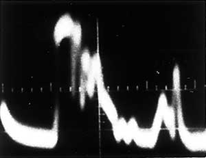
A-mode is a method of displaying echoes acquired in 1 dimension in which depth is represented along 1 axis and an echo amplitude is displayed along a perpendicular axis. The image shown is from the original A-mode study by John Julian Wild, MD, PhD, that revealed the differences in echo pattern between a normal stomach wall and a stomach wall infiltrated by cancer.

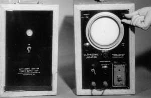
Images show George D. Ludwig, MD, and his first ultrasonic scanning equipment. Dr Ludwig and colleagues conducted experiments for the US Navy in the diagnostic capabilities of ultrasound; they used A-mode presentation of reflected echoes exclusively.
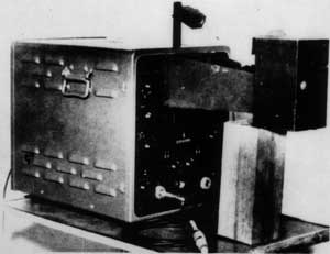
M-mode Imaging (Motion-mode)
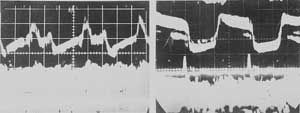
M-mode is a method of display in which tissue interface position is displayed along 1 axis and time is displayed along a second axis. M-mode is used frequently to display echocardiographic data in which heart wall motion and valve motion are displayed as functions of time. The images shown are echo pattern records of the motion of the anterior leaflet of the mitral valve (left, normal; right, stenosis) obtained with the flaw detector that is shown at the bottom of the page.
Flaw detector converted by Inge Edler, MD, and Professor Helmuth Hertz, into an M-mode echocardiographic machine, with their home-built camera attached.