Pre-Seminar Courses

Principles of Sonography and Doppler Ultrasound
Live or internet live activity, up to 4.75 AMA PRA Category 1 Credit(s)™
Wednesday, February 11, 2026, | 6:30 am–12:30 pm
Registration | 6:30 am–5:00 pm | Asbury Rotunda
Breakfast | 6:45 am–7:30 am | Salon 3
Pre-Seminar Location | Grand Harbor, North Ballroom
Overview
Gain a comprehensive understanding of the operating principles of sonography and master their diagnostic applications with enhanced expertise. Explore topics such as the conventional pulse-echo principle, the modern virtual beam-forming principle, and more. Dr. Frederick W. Kremkau is a leading expert on the topic and author of the best-selling book Sonography Principles and Instruments, now in its 10th edition.
Learning Objectives
After participating in this course, participants will have increased knowledge and better competence to:
Explain how ultrasound interacts with tissue.
Demonstrate how transducers work.
Identify how sonographic and Doppler instruments produce displays from echoes.
Recognize and manage common artifacts.
Practice quality assurance and safety.
Faculty
-
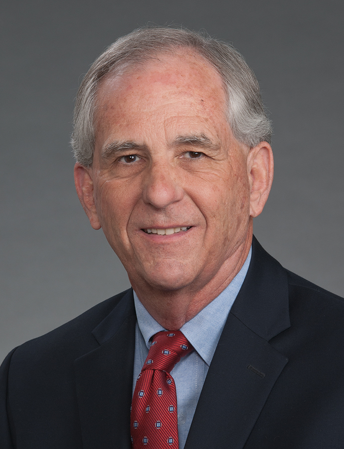
Frederick W. Kremkau, PhD, FACR, FAIMBE, FAIUM, FASA
Professor, Ultrasound Education
Eastern Virginia Medical School, Norfolk, Virginia
Professor Emeritus, Radiologic Sciences
Wake Forest University School of Medicine, Winston-Salem, North Carolina
Agenda
Time | Title | Presenter |
6:30 am–5:00 pm | Onsite Registration Open | Frederick W. Kremkau, PhD, FACR, FAIMBE, FAIUM, FASA |
6:45 am–7:30 am | Breakfast | |
7:30 am–8:15 am | Ultrasound: Sound We Don’t Hear | |
8:15 am–9:00 am | Transducers: Sending and Receiving | |
9:00 am–9:30 am | Instruments: Imaging Anatomy 1 | |
9:30 am–9:45 am | Break | |
9:45 am–10:15 am | Instruments: Imaging Anatomy 2 | |
10:15 am–10:45 am | Artifacts: What Can Go Wrong? | |
10:45 am–11:45 am | Instruments: Imaging and Flow | |
11:45 am–11:55 am | Is it Performing Well? | |
11:55 am–12:15 pm | Is it Safe? | |
12:15 pm–12:30 pm | Questions, Discussions, and Answers | |
12:30 pm | Adjourn |
Agenda subject to change.
The AIUM designates this live activity for a maximum of 4.75 AMA PRA Category 1 Credit(s)™. Physicians should claim only the credit commensurate with the extent of their participation in the activity.
International Ovarian Tumor Analysis (IOTA) Certification Course - Non-CME
In Lieu of CME Credit, this course includes the IOTA certification exam and AIUM certificate of completion.
Wednesday, February 11, 2026 | 1:00 pm–5:30 pm
Registration | 6:30 am–5:00 pm | Asbury Rotunda
Pre-Seminar Location | Grand Harbor, North Ballroom
Overview
Learning Objectives
After participating in this course, participants will have increased knowledge and better competence to:
-
Evaluate an adnexal mass using IOTA and O-RADS models.
-
Obtain IOTA certification (upon passing the exam).
-
Utilize transvaginal ultrasound to discriminate between benign and malignant tumors.
-
Apply new techniques to evaluate patients with presumed endometriosis and or adenomyosis.
-
Identify deep endometriosis to aid in surgical planning with live scanning.
Faculty
-
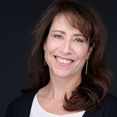
Director of Gynecologic Ultrasound
Diagnostic Ultrasound Associates
Lecturer in Obstetrics, Gynecology and Reproductive Medicine
Harvard Medical School
-
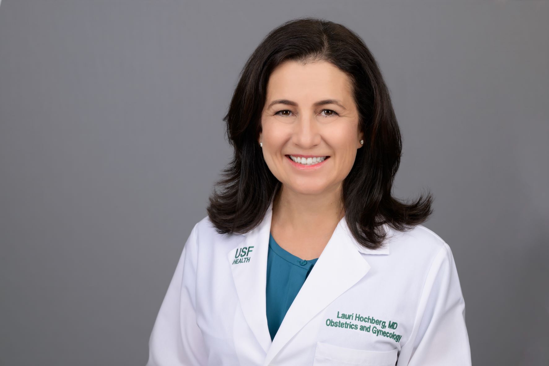
Professor, Division Director of Gynecologic Imaging
Department of OB/GYN
University of South Florida, Morsani College of Medicine
Tampa, FL
-
Assistant Professor Obstetrics and Gynecology
University Hospitals Leuven
Leuven, Belgium
Agenda
|
Time |
Title |
Presenter |
|
1:00 pm–1:10 pm |
Introduction & Overview |
Lauri Silver Hochberg, MD, FACOG |
|
1:10 pm–1:50 pm |
IOTA Terms and Definitions |
Wouter Froyman, MD, PhD |
|
1:50 pm–2:20 pm |
Benign Easy Descriptors and Simple Rules |
Yvette S. Groszmann, MD, MPH, FACOG, FAIUM |
|
2:20 pm–3:20 pm |
IOTA ADNEX Model and 2-step Strategy |
Lauri Silver Hochberg, MD, FACOG |
|
3:20 pm–3:30 pm |
O-RADS |
Yvette S. Groszmann, MD, MPH, FACOG, FAIUM |
|
3:30 pm–3:45 pm |
Break |
|
|
3:45 pm–4:30 pm |
Review and Case Examples |
Yvette S. Groszmann, MD, MPH, FACOG, FAIUM, Lauri Silver Hochberg, MD, FACOG, and Wouter Froyman MD, PhD |
|
4:30 pm–5:30 pm |
IOTA Certification Exam |
|
|
5:30 pm |
Adjourn for the Day |
|
Agenda subject to change. This is a non-CME agenda as of February 2, 2026.
The International Ovarian Tumor Analysis (IOTA) Certification Course, a NON-CME activity, includes the IOTA certification exam and an AIUM Certificate of Completion.


